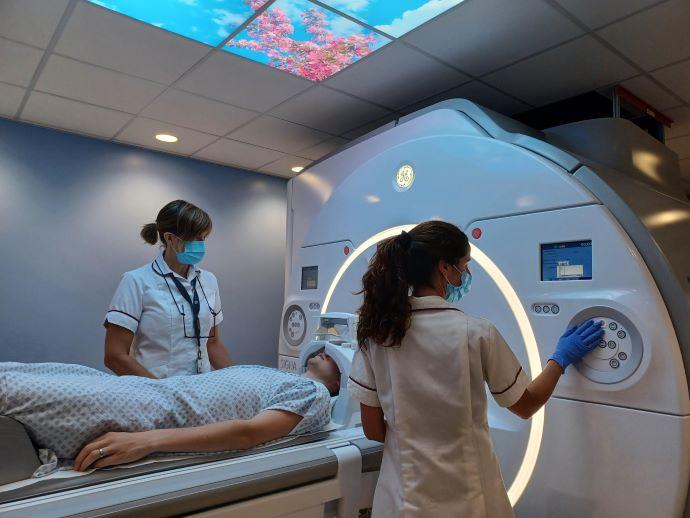
The state-of-the-art 3.0 tesla magnetic resonance imaging (MRI) scanner contains breakthrough AI technology that is used to reconstruct patient images and has twice the strength of the eleven-year old scanner it replaces.
Part of a £2 million upgrade to the hospital’s MRI service, the scanner is reducing waiting times by shortening the length of examinations, including 20% for head imaging. The extra sensitive detection coils and AI technology create an improved signal which allows clinicians to obtain sharper images, helping to diagnose patients with diseases including cancer and bone and joint diseases.
The new kit will also provide opportunities for researchers to develop new methods to detect diseases such as cancer and dementia earlier, leading to earlier diagnosis, and more accurately monitor treatment.
Dr Martin Graves, Professor of MR Physics and honorary consultant clinical scientist at CUH said: “Some patients can become quite anxious during their MRI examination and find it difficult to lie still. This may mean that some of the images obtained using standard imaging techniques are poor quality, but with the use of this new AI-based method we can reduce those scan times making the experience much better for the patient.”
Trained on tens of thousands of high quality images, the AI technology enables the images to be reconstructed more accurately by removing signals which interfere with the images. Addenbrooke’s was the first hospital in the UK to pilot AI technology designed by medical company GE Healthcare. The Trust has now become the first in the country to adopt it with the state-of-the-art 3.0 tesla device.
The scanner also has the capability to image bones by providing X-ray-like pictures but without the X-ray radiation. Dr Andrew Grainger, consultant musculoskeletal radiologist says:
“This new technique is very exciting and produces images like the images of bone that we currently obtain from our CT scanners. In some patients we hope to be able to use this technique to avoid the use of CT and therefore X-ray radiation. In suitable patients it will also allow us to obtain both an MRI and CT equivalent scan at the same time; potentially avoiding the need for two separate hospital visits in patients who previously required a CT and an MRI scan.”
Dr Ferdia Gallagher, Professor of Translational Imaging and honorary consultant radiologist says:
“This new MRI offers a tremendous opportunity to develop cutting edge research in Cambridge. Our researchers are developing novel imaging techniques to detect disease earlier and more accurately, as well methods to image successful response to treatment. These approaches have the potential to improve patient outcome across a range of diseases including many cancers, cardiovascular and neurological disease, and musculoskeletal disorders.”
Image: 3.0 tesla magnetic resonance imaging (MRI) scanner


